


3D imaging is relatively new to the dental industry, but its potential impact is huge. It has the power to revolutionize not only how dentists practice, but also what services and treatments can be offered to patients. This in-depth article explores the potential of 3D imaging and how it can currently support your dental practice.
Over the last decade, the traditional approach to dentistry has shifted from reactive care (i.e., fixing problems that already exist) to preventive care—preventing problems before they show up.
This shift has been driven largely by the development of advanced software, increasing dental practices' efficiency and efficacy. By 2030, the global dental software market will double its 2022 market value, reaching nearly $5 billion, according to data from Grandview Research.
At the forefront of this software boom is imaging technologies, such as 3D imaging. Dentists can diagnose oral health problems much earlier and with greater accuracy and confidence by taking detailed three-dimensional scans of a patient's mouth.
Globally, this software is becoming more accessible and affordable, meaning that even small practices can benefit from the latest innovations.
What Is 3D imaging In dentistry?
In dentistry, 3D imaging is the process of capturing detailed three-dimensional images of a patient's mouth.
3D imaging takes standard dental imaging up a notch—recording cross-sections of the mouth, including a patient's teeth, gums, and other soft tissue. This allows dentists to take detailed scans that clearly reveal areas of decay, gum disease, or other issues—aiding in diagnoses and treatment plans.
In addition to delivering precise information about the anatomical structure of a patient's teeth and other oral components, 3D imaging offers several improvements over its two-dimensional counterpart.
These include:
- More accurate diagnosis for common oral infections
- Improved prognosis for existing treatment plans
- Greater transparency in patient interactions due to more detailed assessments
- Enhanced surgical outcomes through pre-operative planning
- Ability to perform minimally invasive procedures
- Improved treatment outcomes with advanced 3D analysis
3D imaging technology is used primarily in implantology and orthodontics to create detailed models of the jawbone structure and its relationship with other facial structures. With this data, clinicians can make more informed decisions regarding treatment plans.
Cone Beam Computed Tomography (CBCT) is dentistry's most common 3D imaging technology. It can produce high-resolution images considerably faster than traditional CT scans. CBCT offers quick scans with minimal radiation exposure while capturing highly detailed images that can be used for diagnosis, treatment planning, and post-surgery evaluation.
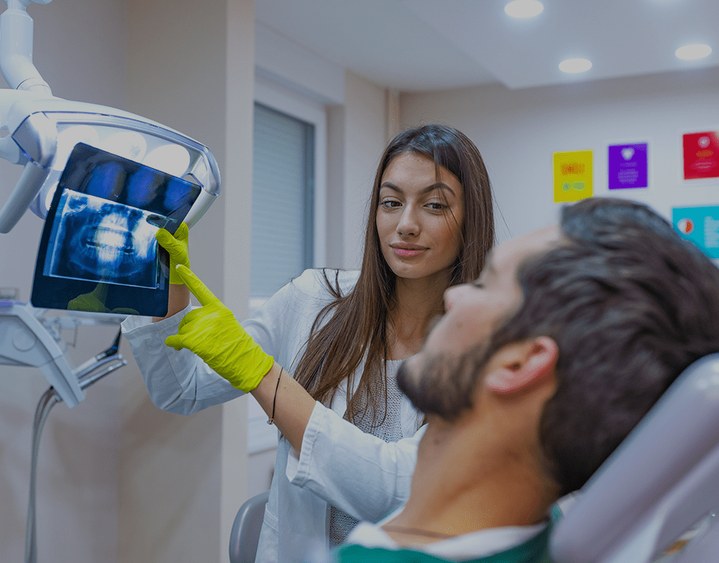
What To Expect During A CBCT Scan
When you picture a CBCT scan, you might think of a clunky, ominous machine like those found in hospitals. In reality, CBCT equipment is approachable and efficient. It requires no preparation or anesthesia and can be done in seconds.
Here's what to expect from a CBCT scan:
-
Before the scan: Your dentist may take X-rays prior to scanning to get detailed information about your oral anatomy. You may also be asked to fill out a questionnaire about any allergies you may have or previous medical conditions that could affect the scan results.
-
During the session: The whole procedure only takes a few minutes. The dentist will seat you upright on an armchair as the machine rotates around you, taking 3D photos of your face and jawbone. To ensure accuracy, you will need to remain still throughout the entire scan, as even minor head movements can interfere with its accuracy.
- Afterward: The computer usually takes around ten minutes to process your photos. Once your scans are complete, your dentist will review the images and talk with you about what they mean in the context of your treatment plan.
How Does 3D Dental Scanning Work?
3D imaging sounds like a complicated and technical process. But it's a lot easier than you think.
At its core, all 3D imaging does is capture two-dimensional images from different angles. The CBCT scanner works by spinning around the patient and capturing hundreds of images in rapid succession.
These images are then combined together to create detailed three-dimensional models with several times greater resolution than traditional two-dimensional (2D) x-rays can offer.
This level of detail is invaluable when diagnosing conditions or examining fine details such as tooth fractures or black dots on teeth, which are often overlooked on 2D scans.
3D Dental Imaging: Benefits Of 3D Technology
3D imaging is increasingly becoming dentistry's gold standard for diagnosis, treatment planning, and monitoring. Here are six ways your practice can leverage this technology:
1. More Reviewable Information For Diagnostics
Two-dimensional representations of a patient's mouth are helpful for certain procedures, but they are also limited in their ability to show elements like tooth orientation and root structure.
Adding a third dimension to dental imaging offers a much better perspective of your patients' oral conditions. The CBCT scanner provides clear and detailed images without any distortion, which helps you make quick and accurate diagnoses.
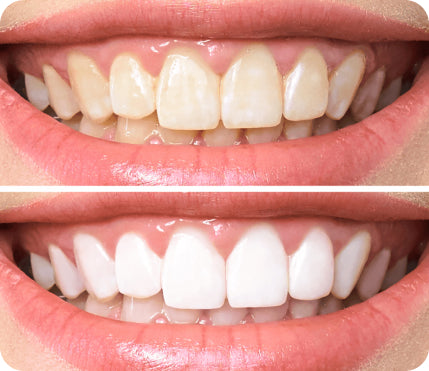
When diagnosing pathologies, planning for dental implant surgery, and performing in-office teeth whitening, 3D imaging offers a more comprehensive view of the patient's teeth and jaw.
For patients with complex dental problems, 3D imaging creates a bird's-eye view of their condition, which is invaluable when deciding the next course of action.
2. Improved Visualizations
Cone beam imaging creates detailed 3D models of the patient's mouth, allowing clinicians to visualize a wide range of complex dental issues. 3D imaging splits the jaw into multiple sections, which makes it easier to pinpoint problems like cavities and periodontal disease.
Cone beam visualizations now play an essential role in a wide range of oral procedures:
- Endodontics, including root canal treatments and apical surgeries
- Tooth extraction
- Veneers, dental implants, and crowns
- TMJ disorder diagnosis and treatment
- Orthodontic procedures such as braces and Invisalign
In addition to giving clinicians a more detailed picture of the patient's teeth, 3D imaging also helps them identify any possible complications before they occur.
Impacted wisdom teeth are perfect examples of this. By scanning the patient's jaw and viewing the results in 3D, dentists can determine how their surgical movements will affect the surrounding structures, reducing the risk of damaging essential nerves during the procedure.
3. Better Surgical Planning
Oral surgery is ultimately an error-prone process. Using cone beam imaging, dentists and oral surgeons can more accurately prepare for upcoming surgeries.
The data captured by the CBCT scanner can help surgeons determine the size, shape, and depth of anatomical structures such as jawbones and sinuses, which is invaluable information when planning an operation.
For example, a patient with a missing posterior tooth that needs an implant would normally have to go through a laborious process of manually measuring with calipers.
For the patient, this is cumbersome and uncomfortable. For the clinician, it entails a certain degree of guesswork on the day of the procedure.
With 3D imaging, the patient can visit an imaging center, spend 15 minutes having their mouth captured, and get a detailed 3D model showing all the relevant measurements.
This model can then be sent to the oral surgeon, who will use it to plan the implant surgery more accurately.
4. Reduced Radiation Exposure
Several studies have linked dental radiation to increased risks of brain, breast, and thyroid cancers. CBCT scanners deliver significantly less radiation than traditional CT scans, making them a much safer option for patients.
Particularly for dental procedures that require multiple scans over time (such as braces treatment and teeth retainers) and those that require wide-area imaging, 3D imaging technology can significantly reduce the amount of radiation to which a patient is exposed.
With traditional CT scans, the patient is exposed to a high dose of radiation for an extended period of time—a benefit especially critical for young patients, pregnant women, and other groups sensitive to radiation exposure.
5. Improved Communication With Patients
Put yourself in your patients' shoes: Does waiting several weeks for a detailed prognosis or undergoing several tests with conventional imaging sound like a pleasant experience?
And if a patient has a severe problem like broken teeth, a fungal infection, or a cavity, they are probably worried about what the following steps will look like.
3D models are generated much more quickly than traditional diagnostic tools and are easier to understand. This makes them an invaluable educational tool for dentists when communicating problems to their patients efficiently and comprehensibly.
Patients who get the information they need quickly are less anxious about their treatment. And the improved transparency in the process makes you more trustworthy and credible in their eyes.
6. Reduced Margin Of Error
Like every medical profession, oral care is performed by humans (for the most part). This entails some level of risk, as humans are prone to making mistakes.
3D imaging helps reduce the margin of error by providing dentists with detailed and accurate images of their patients' teeth, jawbones, and surrounding tissues.
Root canals, dental implants, and orthodontic surgeries all require precise planning, and 3D imaging helps surgeons plan their course of action far ahead of the actual procedure. This reduces the chances of mistakes and gives your patients more peace of mind when undergoing complex operations.
Common Uses Of CBCT
Cone beam imaging is used in a wide range of dental procedures, from diagnosing oral diseases to planning orthodontic and restorative treatments.
1. Assessing Trauma To Teeth, Jaws, And Other Oral Structures
The human mouth is complex, and several factors go into treatment planning for chipped teeth, broken jaws, and other traumas.
- The rate at which a patient's existing oral infrastructure is shifting affects whether or not a permanent surgical procedure suits them.
- Trauma from impact can have underlying effects on the mouth as a whole, such as damage to adjacent teeth, bones, and soft tissues.
- The anatomy of adjacent structures (i.e., teeth, bones, and gums) can determine whether a procedure is safe or possible.
- Underlying problems with the dimensions and structure of the mouth may rule out adjunctive treatments.
With two-dimensional representations of a patient's mouth, it can be difficult to accurately assess their overall oral condition. Small but critical factors like the exact location and size of a microfracture that was residually caused by trauma may be overlooked without 3D imaging.
2. Identifying Abnormalities In Wisdom Teeth Growth
85% of people have their wisdom teeth removed. Third molar removal is generally considered a "must" for young adults, and two-dimensional imaging often suggests that wisdom teeth removal is the most viable solution.
But wisdom teeth don't always need to come out. And a proper analysis of protruding wisdom teeth with cone beam imaging could save thousands of patients the pain and cost of a wisdom teeth removal procedure.
For those who need wisdom teeth removal, CBCT can provide greater transparency into the procedure at an earlier stage. This helps patients plan further in advance and helps dentists plan their course of action to minimize risks and complications.
3. Making Sense Of Unusual Oral Symptoms
The human mouth is connected to the respiratory, gastrointestinal, and immune systems. Certain oral diseases can be indicative of serious underlying problems in other parts of the body.
Diagnosing an illness in another part of the body often begins with a comprehensive assessment of dental symptoms like bone abnormalities or swelling.
3D imaging provides an opportunity to inspect the precise location of bone and tissue abnormalities, helping to identify the source of discomfort. This helps dentists determine whether these indicate more significant problems that require further medical attention or evaluation.
4. Planning For Dental Implants
Dental implants require detailed planning and precise calculations to ensure that they fit comfortably in the mouth and don't cause any damage to existing teeth or gums.
Cone beam imaging allows dentists to assess a patient's mouth's size, shape, and positioning before implantation. This helps them plan for a successful procedure with minimal risks and maximum comfort for their patients.
Aside from preparation and planning, CBCT imaging can also be used to check for any potential issues after the dental implant procedure, ensuring the implant fits properly long-term.
5. Endodontic Procedures
Endodontic procedures involve working on the pulp of a tooth, which can be difficult to assess without 3D imaging. CBCT imaging is heavily used in endodontics, as it helps dentists to identify cracks or canals that may have eluded detection in two-dimensional imaging.
CBCT provides a 3D view of anatomical features that can't be seen with traditional 2D imaging techniques, such as intraoral, panoramic, and other modalities. This allows dentists to more accurately assess the condition of a tooth's root and plan a treatment accordingly.
Wrapping Up
3D imaging is revolutionizing dental practices, providing practitioners with a more accurate way to diagnose and treat their patients. It significantly reduces the risks associated with dental procedures and allows dentists to plan treatments more effectively.
For patients, these benefits mean safer and more comfortable treatments with greater satisfaction.
Want to learn more? Here are the questions our customers ask us the most.
Do You Need To Drink Water For A 3D Scan?
Unlike ultrasounds, 3D scans do not require you to drink any special fluids. Water does not play a significant role in the results of the scan, and the 3D scanning process is only around 10 to 15 minutes long, so patients can typically wait until after the scan to drink water.
Is A 3D Dental Scan Necessary?
A 3D dental scan is usually only necessary for the following cases:
- Oral trauma that requires a more detailed scan to evaluate the severity of the injury
- Wisdom teeth extractions or other tooth removal procedures that require precise measurements
- Endodontic procedures that require 3D imaging for a more accurate diagnosis
- Prognosis for severe, time-sensitive oral health conditions like cancer and cysts
- Dental checkups assessing wisdom teeth growth and impacted teeth
- Dental implant planning and execution
How Long Does A 3D Teeth Scan Take?
3D teeth scans generally take no more than 15 minutes to complete. This is about as long as a regular panoramic X-ray, but with much higher accuracy. The resulting images are also much more detailed and easier to analyze than 2D scans.
Which Type Of Dental Radiograph Shows 3D Images?
Cone beam computed tomography (CBCT) is the most common type of imaging used to create 3D images. This type of radiograph produces high-resolution scans that can be viewed from multiple angles, providing a comprehensive view of the patient's oral anatomy It combines multiple two-dimensional images into a single three-dimensional one to create an accurate picture of the teeth and jaw.
How Much Does A 3D Dental Scan Cost?
3D dental scanning generally costs between $350 and $600 per session, depending on the type of scan, the location, and the practitioner. Some insurance plans cover 3D imaging for certain procedures, but it's always best to check with your insurer before making an appointment.